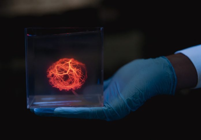
Stanford researchers have developed a new method to design and 3D print vascular systems, marking a major step toward producing transplantable organs from a patient’s own cells and potentially reducing wait times and rejection risks for thousands on US transplant lists.
The research, published on 12 June in Science, demonstrates an advance in regenerative medicine by enabling faster, more precise generation of the complex networks of blood vessels needed to support lab-grown organs.
According to the Stanford team, their new platform generates vascular designs at speeds approximately 200 times faster than previous methods and produces results that closely resemble the intricate architecture of native human tissues.
“The ability to scale up bioprinted tissues is currently limited by the ability to generate vasculature for them – you can’t scale up these tissues without providing a blood supply,” said Alison Marsden, professor of cardiovascular diseases, pediatrics, and bioengineering at Stanford, and co-senior author of the paper.
The new algorithm developed by the Stanford team not only produces detailed vascular trees but also ensures realistic fluid dynamics, even distribution of blood, and a complete closed-loop system with a single inlet and outlet.
Zachary Sexton, a postdoctoral scholar in Marsden’s lab and co-first author of the paper, said the algorithm allowed them to generate a model for a human heart’s vascular tree containing one million vessels in just five hours.
“That task hadn’t been done before, and probably would have taken months with previous algorithms,” Sexton said.
While 3D printers are not yet capable of fabricating networks with that level of detail, the team printed a vascular model with 500 branches and tested a simplified version that kept human embryonic kidney cells alive.
Using a 3D bioprinter, they created a thick tissue ring with a 25-vessel network and demonstrated that a nutrient-rich fluid pumped through the system could sustain the surrounding cells.
“We show these vessels can be designed, printed, and can keep cells alive,” said Mark Skylar-Scott, assistant professor of bioengineering at Stanford and co-senior author of the study.
“We know that there’s work to do to speed up the printing, but we now have this pipeline to generate different vascular trees very efficiently and create a set of instructions to print them.”
The researchers caution that these printed vessels are not yet true, functional blood vessels. They lack the muscle and endothelial cells required to mimic real vascular systems. However, the team sees this as a foundational step toward that goal.
“This is the first step toward generating really complex vascular networks,” said Dominic Rütsche, co-first author and postdoctoral scholar in Skylar-Scott’s lab. “We can print them at never-before-seen complexities, but they are not yet fully physiological vessels. We’re working on that.”
Stanford researchers are now integrating their vascular models with lab-grown human heart cells, aiming to build a full-scale, functional bioprinted heart.
According to Skylar-Scott, the team has already generated enough heart cells from human stem cells and is actively combining them with the vascular designs to advance the field of personalized organ fabrication.
“This is a critical step in the process,” Skylar-Scott said. “We are now actively putting the two together: cells and vasculature, at organ scale.”


