


A recent collaboration between researchers from the California NanoSystems Institute at UCLA, and researchers from Stockholm and Uppsala University and SciLifeLab in Stockholm, has resulted in the creation of an innovative diagnostics tool that can identify cancerous tumours and infections like tuberculosis.
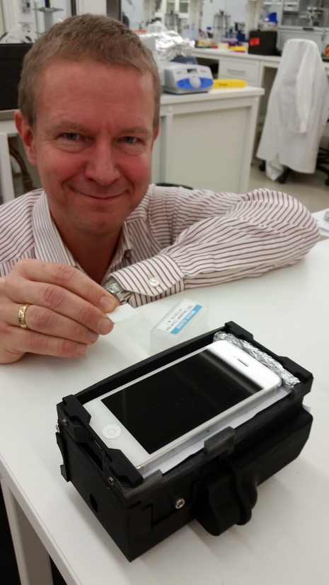
The invention, a new smartphone-based microscope produced with a 3D printer, can be used to analyse samples of tumours or bacteria, virus and fungal cells, while also serving as a potent weapon in the battle against antibiotic resistance.
Mats Nilsson from Stockholm and Uppsala University and SciLifeLab in Stockholm, said the microscope and smartphone can show how the DNA chains in the sample look like, adding that if the doctors detect certain variants, they will know exactly what form of cancer, bacteria or virus is involved.
“I am used to big, expensive and complicated machines which fill a whole room to do the DNA sequencing,” Mr Nilsson said.
“To me, the idea to use a smartphone for this was very interesting. This opens the way to many new and very important fields of application.”
He said the little 3D-printed microscope could be of great significance for many people, including those in poorer parts of the world, who can’t afford the advanced, lab-based testing which is only performed at major hospitals.
According to him, if produced in large quantities, the new device could be manufactured for less than $500. It could run on the smartphone battery, independent from constant power supply.
“Antibiotics are effective against bacteria. But now we are losing that weapon when bacteria become resistant. When it comes to tuberculosis, antibiotic resistance is a big problem,” Mr Nilsson continued.
“However, if we could look at the DNA-level and find out if a bacterium is sensitive to a certain type of antibiotics, we could choose the right treatment from the very beginning. This is where I think this concept has its great strength.”
The technique was already used to identify cancerous tumours in the colon, allowing doctors to rule out the ineffective treatments by analysing certain mutations in the tumour.
It also allows for a quick transfer of images and information about DNA to a doctor located in a different part of the world, and researchers are also hopeful that the technology can facilitate the diagnosis of viral infections like Ebola or Zika virus.
The microscope was developed by researchers from the California NanoSystems Institute at UCLA, while their peers from Sweden worked with the analysis of the DNA chains.


